
Electron Microscope
Best for:
Desktop wallpapersArt printsSocial media postsEducational contentPoster printsScience communication
Sample prompt used to generate the hero image
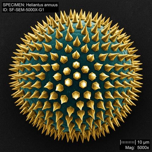
Scanning electron microscope (SEM) image of a single pollen grain at 5000x magnification, 1:1 square format. The pollen grain's surface is revealed as an elaborate alien architecture — spikes, ridges, and pore openings creating a complex three-dimensional topography. Monochromatic grey-scale with dramatic directional lighting from the upper right creating strong shadows that emphasize every surface detail. The depth of field is characteristic of SEM — nearly everything is sharp. The background fades to a smooth dark void where the electron beam didn't reach. Scientific specimen aesthetic with a scale bar in the lower right showing "5 μm."Prompt Variations
Section titled “Prompt Variations”SEM image of [SPECIMEN — e.g., insect compound eye / butterfly wing scale], [MAGNIFICATION], grayscale, directional lighting, three-dimensional topology, scale bar, [FORMAT].SEM image of [MATERIAL — e.g., fabric fiber weave / metal fracture surface / crystal growth], [MAGNIFICATION], grayscale, surface detail, dramatic shadows, scale bar, [FORMAT].SEM image of [FOOD — e.g., sugar crystals / bread crumb cross-section / coffee ground], [MAGNIFICATION], revealing alien microscale geometry, grayscale, directional lighting, [FORMAT].False-color SEM image of [SPECIMEN], scientific colorization applied — [COLORS assigned to different elements/regions], dramatic detail, scale bar, [FORMAT].Split-image showing [OBJECT] at normal scale on the left and SEM magnification on the right, revealing the hidden micro-structure, grayscale SEM half, scale bars, [FORMAT].Create a scanning electron microscope (SEM) image of [SPECIMEN — e.g., an insect compound eye] at [MAGNIFICATION — e.g., 5000x] magnification. Monochromatic grayscale with dramatic directional lighting from the upper right, revealing elaborate three-dimensional surface topology. Nearly infinite depth of field, dark void background, and a scale bar in the lower right. Scientific specimen aesthetic. 1:1 square format.Create an SEM image of [MATERIAL — e.g., fabric fiber weave or a metal fracture surface] at extreme magnification — grayscale, strong directional shadows emphasizing surface detail, alien microscale geometry, scale bar in the corner. 16:9 landscape format.Create a false-color SEM image of [SPECIMEN] with scientific colorization — [COLORS] assigned to different structures — over dramatic grayscale topology, directional lighting, dark void background, scale bar. 16:9 landscape format.scanning electron microscope image of [SPECIMEN], [MAGNIFICATION] magnification, monochromatic grayscale, dramatic directional lighting, three-dimensional surface topology, infinite depth of field, dark void background, scale bar --ar 1:1 --s 100SEM image of [MATERIAL] surface, extreme magnification, grayscale, strong directional shadows, alien microscale geometry, scientific specimen, scale bar --ar 16:9 --s 100false-color scanning electron microscope image of [SPECIMEN], scientific colorization, [COLORS], dramatic surface topology, directional lighting, dark background, scale bar --ar 16:9 --s 150scanning electron microscope image, [SPECIMEN], extreme magnification, monochromatic grayscale, dramatic directional lighting, three-dimensional topology, infinite depth of field, dark void background, scale bar, scientific specimenNegative Prompt
color photograph, blurry, shallow depth of field, artistic painting, low qualityfalse-color SEM image, [SPECIMEN], scientific colorization, [COLORS], dramatic surface topology, directional lighting, dark background, scale barNegative Prompt
natural photograph, oversaturated rainbow, painting, blurImage-to-Image Transformations
Section titled “Image-to-Image Transformations”Using the attached photo, reimagine it as if viewed through a scanning electron microscope at extreme magnification. Convert to grayscale. Reveal hidden three-dimensional surface topology invisible at normal scale. Add dramatic directional lighting from the upper right. Nearly infinite depth of field. Dark void background. Add a scale bar in the lower right. Scientific specimen aesthetic.[Upload Photo] "Reimagine this photo as a scanning electron microscope image — grayscale, extreme magnification revealing 3D surface topology, dramatic directional lighting, infinite depth of field, dark void background, and a scale bar."[IMAGE_URL] scanning electron microscope image, grayscale, extreme magnification, dramatic directional lighting, surface topology, dark void background, scale bar --iw 1 --ar 1:1Tips & Best Practices
Section titled “Tips & Best Practices”- Grayscale only (unless false-color): Real SEM uses electrons, not light, so images are monochromatic.
- Dramatic directional lighting: “Lighting from upper right” casting strong shadows that reveal 3D surface features.
- Name the magnification: “5000x magnification” calibrates the level of detail and alien-ness.
- Scale bar: “Scale bar showing ‘5 μm’” adds scientific authenticity.
- Pairs well with: X-Ray / Medical Imaging (both reveal hidden structures), Cinematic Macro Photography (next step down in magnification)